A dermatology exam is a detailed medical assessment of your skin, hair, and nails performed by a board-certified dermatologist to detect early signs of disease or damage.
During a derm exam, the specialist examines your entire body from head to toe, identifying unusual spots, moles, or growths that may indicate a higher skin cancer risk.
This process supports early detection of skin cancer and other conditions, allowing for timely and effective treatment. At DermOnDemand, every dermatology exam is reviewed by experts led by Dr. Hannah Kopelman, ensuring accurate, confidential, and personalized care for every patient.
Key Takeaways
- A dermatology exam is a complete skin evaluation performed by a board-certified dermatologist to identify unusual spots, moles, or lesions that could indicate early signs of disease.
- During a derm exam, the specialist inspects the body from head to toe, including less visible areas, to ensure early detection of skin cancer and other conditions.
- Preparing for your visit by removing makeup or nail polish and noting any new or changing spots helps your dermatologist evaluate your skin more accurately.
- Regular skin checks—both at home and with a dermatologist—are essential for reducing skin cancer risk, especially for people with a higher risk or fair skin type.
- At DermOnDemand, Dr. Hannah Kopelman and her team provide expert, discreet dermatology examinations designed for precision, comfort, and proactive prevention.
What Happens During a Dermatology Exam
A dermatology examination is a clinical evaluation of your skin, hair, and nails to identify unusual spots, moles, or growths. It’s performed by a dermatologist trained to assess different types of skin and recognize early warning signs of disease.
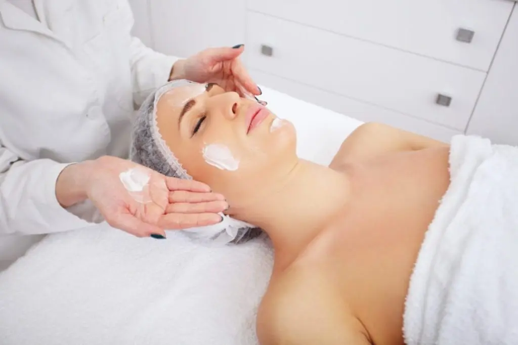
During a derm exam, the doctor looks at your skin from head to toe. The process is simple and noninvasive. You’ll be asked to remove clothing, and a gown is provided to maintain comfort and privacy. Each area of skin is examined for color, shape, and texture changes.
A dermatology exam can take 10 to 20 minutes, depending on the number of lesions or moles. Areas like the scalp, soles, and nails are checked carefully because skin cancer risk can extend beyond sun-exposed regions.
For complete accuracy, dermatologists may also examine private areas if there are visible lesions or symptoms.
If you’ve never had a derm exam, it’s best to schedule one when you notice new or changing moles, or if you have a higher risk due to family history, fair skin type, or extensive sun exposure. Most experts recommend annual skin checks as part of preventive care.
Derm Exam Steps and Procedure
Preparing for Your Exam (Patient Tips)
Preparing for your exam is simple: avoid makeup, nail polish, or tinted lotions that may cover your skin’s surface. Bring a list of medications and note any new spots or areas of concern.
Step-by-Step Derm Exam Process
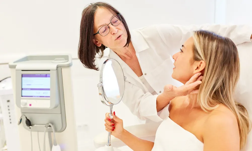
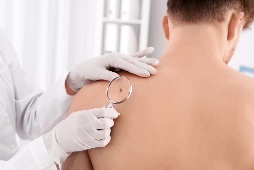
The dermatologist begins with a visual inspection. Each area is evaluated for size, symmetry, and color. Lesions are documented using digital photography or diagrams. If needed, the doctor may use a dermatoscope – a small magnifier with light – to examine deeper layers of the skin.
What the Full-Body Exam Includes
Key steps in a dermatology exam include:
- Visual review of the entire body surface.
- Palpation to check texture, firmness, or tenderness.
- Mapping of moles or marks for follow-up comparison.
- Evaluation of hair and nails for signs of systemic conditions.
A full body exam ensures that hidden or subtle changes are not missed, improving early detection of skin cancer and other dermatologic conditions.
Understanding Derm Exam Findings and Terms
Common Lesion Types Explained
Dermatologists classify findings based on lesion type – for example, macules (flat spots), papules (small bumps), or plaques (larger raised areas). Knowing these terms helps patients understand their reports after a dermatology examination.
Documentation and Photography Best Practices
Every skin exam involves documentation. Photos or detailed notes allow comparison during future visits, especially if you’re monitoring moles or unusual spots. Consistent record-keeping helps identify subtle changes over time, a key factor in diagnosing early melanoma or other conditions at higher risk of progression.
Red Flags and When to Seek Care

Warning Signs to Watch For
Some changes deserve immediate attention. You should check your skin regularly and contact a dermatologist if you notice:
- New or fast-growing moles.
- Changes in shape, border, or color.
- Itching, bleeding, or pain in an existing spot.
These signs can indicate early detection of skin cancer, especially in patients with a family history or fair skin.
When to Schedule Follow-Ups
If your dermatologist identifies suspicious lesions, a biopsy may be recommended. Routine follow-ups are usually scheduled every 6 to 12 months, depending on your risk profile and number of moles.
Regular skin checks are one of the most effective ways to protect against advanced disease. The goal is not only diagnosis but prevention – recognizing abnormal cells before they become dangerous.
Expert Tips from Dermatologists
Professional Guidance from Dr. Hannah Kopelman
Board-certified specialists like Dr. Hannah Kopelman emphasize that prevention starts with awareness. Patients should perform monthly self-checks at home, noting any new or changing areas. A simple mirror inspection from head to toe helps you detect changes early.
Why Professional Exams Matter
Professional exams deliver greater accuracy because dermatologists use advanced diagnostic tools and understand subtle variations that may signal disease. Combining at-home checks with expert care reduces your long-term skin cancer risk and supports healthy skin for life.