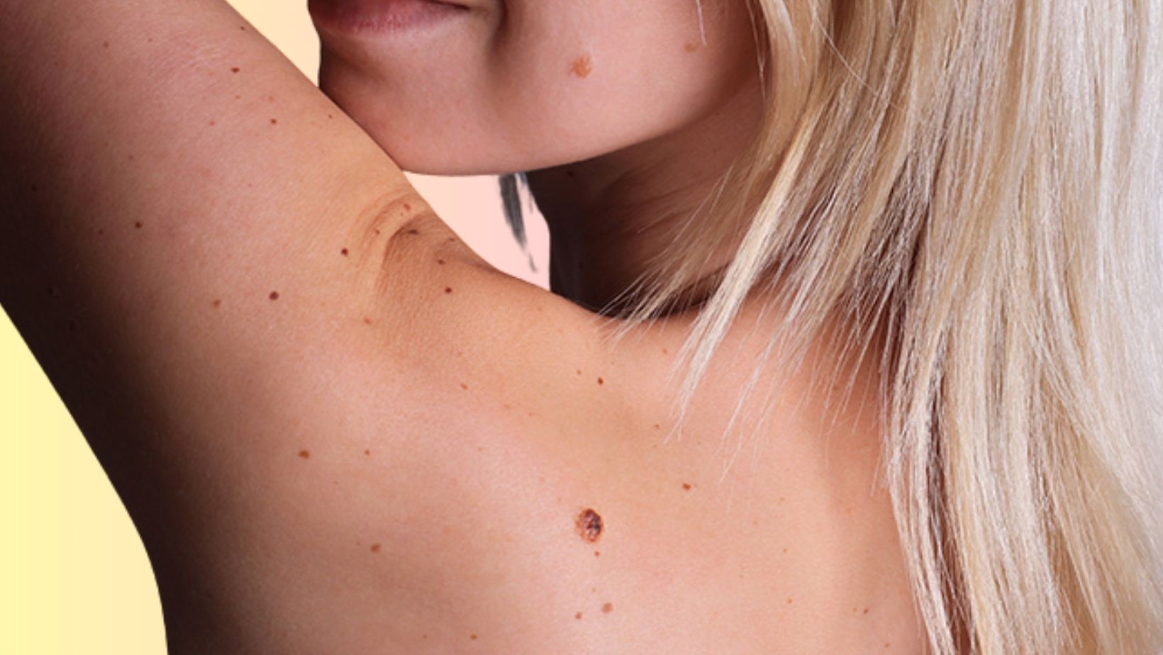
You may have stumbled upon a mole on your skin that doesn’t quite fit the standard image—perhaps it’s flesh-colored or less pigmented than others. Chances are, this is an intradermal pigmented nevus, a benign lesion where melanocytes have nestled themselves deeper within your skin’s layers. Their shape and size can be varied, some presenting as small, dome-shaped bumps, while others may be flatter or possess a tiny stalk. Typically showing up on the neck, head, or trunk, these nevi are nothing to be alarmed about. For those who’ve spotted such a growth, knowing that these are common and benign can be comforting, though keeping an eye on them for any changes remains important.
Despite their benign nature, the appearance of an intradermal pigmented nevus can cause unease, particularly when they manifest in visible areas or in large numbers. It is crucial to understand that these are common skin occurrences and that their presence is typically harmless. Nevertheless, any changes in these nevi should be observed, as they provide an important visual indicator of skin health.
Intradermal Nevus Causes
Intradermal nevi arise from a confluence of genetic elements and environmental influences. A hereditary propensity for moles can mean a higher likelihood of developing intradermal nevi. While sunlight is a known catalyst for various skin lesions, intradermal nevi are not as directly implicated by UV exposure as other mole types. However, they can still be affected by significant sun exposure.
Hormonal changes are another contributing factor, with life events such as puberty and pregnancy often correlating with changes in mole appearance or the development of new growths. This section aims to delve into the potential genetic predispositions and environmental triggers that may increase the occurrence of intradermal nevi, as well as to discuss the impact of hormonal fluctuations on these skin growths.
Diagnosis and Identification
The identification of an intradermal nevus is typically achieved through clinical examination. A dermatologist can assess the mole’s shape, size, color, and other characteristics to distinguish it from other skin lesions. You can do a preliminary teledermatology consult to evaluate whether you have this benign tag. When a nevus presents atypical features, or if there’s a change in its appearance, a skin biopsy might need to be performed. This is a precautionary measure to ensure that the growth is benign.
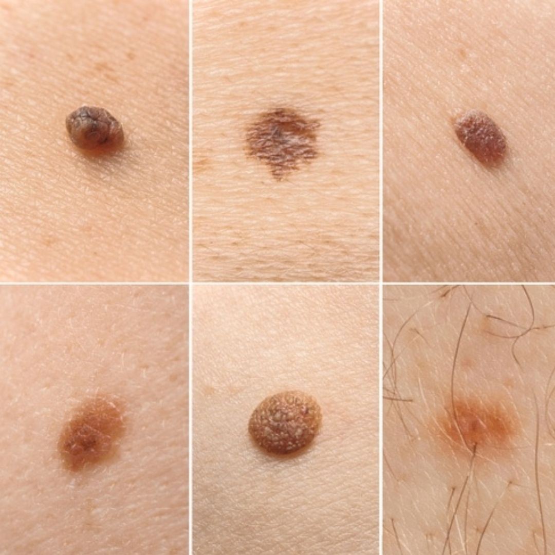
A biopsy entails the surgical removal of a portion of the lesion, which is then examined microscopically for signs of malignancy. While this may sound daunting, it is a routine procedure that can provide peace of mind. In this section, we explain the diagnostic process for an intradermal nevus and emphasize the importance of monitoring for any changes that may warrant a more thorough examination.
Treatment and Management
Intradermal nevi, in most cases, do not require medical treatment. They are generally benign and only removed if they cause discomfort or for cosmetic reasons. If removal is desired, surgical excision is a common approach where the nevus is carefully cut out. Laser treatment offers a non-surgical alternative, although it may not be suitable for all types of nevi and could require multiple sessions to be effective.
It’s essential for individuals to discuss potential treatment options with a healthcare provider, considering factors such as the size and location of the nevus, as well as personal health history. This section discusses the various treatment approaches for intradermal nevi, when treatment might be necessary, and what to expect from the removal procedures.
Potential for Malignancy
Although intradermal nevi are largely benign, they are not without risk. It’s essential for individuals to monitor their moles for any changes in size, shape, or color, which could indicate a potential for malignancy. New growths or significant changes to existing nevi should be evaluated by a healthcare professional. For those with a high number of moles or a family history of skin cancer, regular dermatological check-ups are recommended.
This section emphasizes the importance of self-monitoring and professional skin evaluations as preventative measures. By staying informed and vigilant about their skin growths, individuals can effectively manage their risk and detect potential problems early.
Intradermal nevi are a frequent dermatological finding and are typically noncancerous. Their exact cause is not fully understood, but they are influenced by a combination of genetic makeup and environmental factors, with hormonal changes also playing a role. While diagnosis is usually straightforward, it is critical for individuals to monitor their skin for any changes and consult with a dermatologist as needed. The key to managing intradermal nevi is awareness and proactive monitoring to ensure that any potential risks are identified and addressed promptly. Through understanding and attentive care, individuals can maintain healthy skin while keeping an eye on any developments related to their intradermal pigmented nevus.


